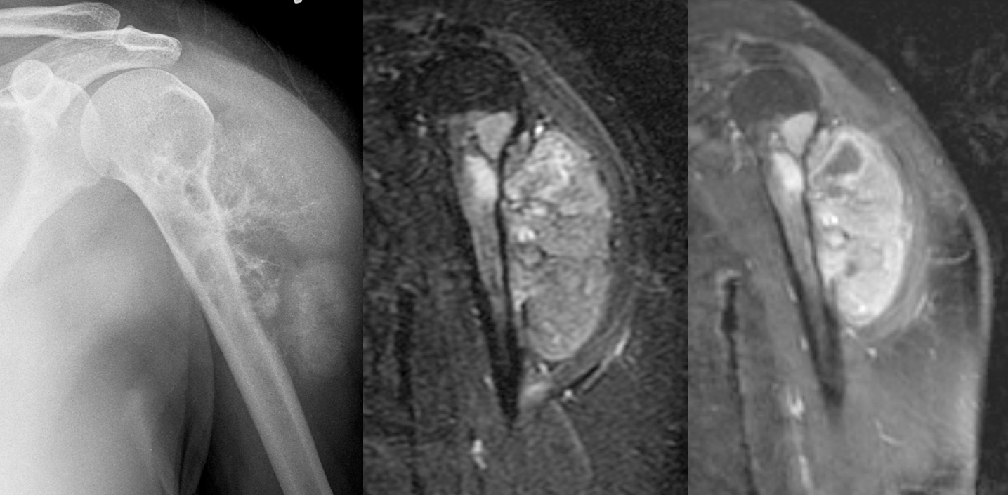
[Left]: X-ray shows a mass arising from the left proximal humerus and extending into the adjacent shoulder soft tissues with really aggressive periosteal reaction ("hair on end"). The proximal humerus itself is also heterogeneous with lucent areas. The lateral surface of the upper humerus shows "saucerization," where the cortex is thinned out and looks like a saucer seen on edge.
[Middle]: MRI IR sequence shows a hyperintense bony mass with large soft tissue component.
[Right]: MRI postcontrast T1 IDEAL shows that the mass is enhancing.
This turned out to be high-grade surface osteosarcoma.
I find these posts fascinating, but I am a layperson with only whatever medical knowledge a doctor father who loved to share imparted. I can somewhat understand these images, and the MRI looks obviously anomalous to me, but I always struggle with X-rays! Would anyone care to help me understand what exactly it is in the X-ray that indicates an issue? To my uneducated eyes, it doesn’t seem that different from a healthy shoulder.
Is it the fact that there’s “cloudiness” between the bone and skin? I guess I can somewhat pick that out now, but I just don’t feel confident I would ever have noticed it on my own without the context of this post.